


Acknowledge Core
Proper Citation:
University of Florida ICBR Cytometry Core Facility, RRID:SCR_019119
Contact
ICBR-Cytometry@ad.ufl.edu
(352) 273-6032
Locations
Cancer & Genetics Research Complex
Room 292 CGRC
2033 Mowry Road
Gainesville FL 32610


The Cytometry core offers project consulting, experimental design and optimization as well as data analysis services to help researchers with large-scale and small-scale experiments acquired on our sorters and cytometry instruments. We coordinate closely with other ICBR cores (Next-Gen Sequencing, Gene Expression, Bioinformatics, Monoclonal Antibody, Electron Microscopy and Proteomics) on shared projects. Our core offers a full suite of services from staff assisted services to training courses for all users, as well as instrument training for 24/7 access to our self-service equipment usage.
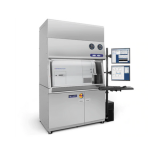
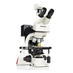
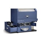
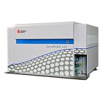
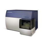
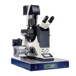
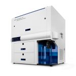
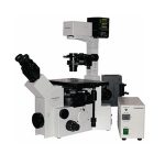
The Flow Cytometry core provides a variety of tools and staff expertise for both live and fixed cell analysis. The laboratory incorporates numerous flow cytometers, from simple entry-level instruments to high-end 5-laser, 17-parameter instruments as well as a spectral cytometer to analyze your cells. We have 3 FACS ARIA’s and a SONY SH800 sorters to help users sort your cells of interest. In addition, the laboratory offers a suite of microscopy equipment for self-service users. The microscopes are well equipped with a variety of excitation lines from near UV to red and can collect a variety of emission colors (depending on dye combinations), with viable cell time lapse and extensive computed parameter capabilities, including FRET, FRAP, image stitching, and 3D reconstruction. We offer a host of Microscope brands from a Leica SP5 Confocal, a Nikon Multiphoton Confocal system, a Nikon Live cell Imaging platform, 2 Olympus spinning Disk Confocal instruments, as well as our new Keyence BZX800.
 UNIVERSITY OF FLORIDA SCIENTIST TO FLY ON BLUE ORIGIN SUBORBITAL MISSION

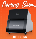
 Advancing Spatial Transcriptomics Research with 10x Genomics Visium CytAssist
 ICBR Contributes to Unveiling Regulatory and Stress-Responsive Networks in Passion Fruit

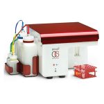
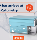
 Introducing the NanoFCM NanoAnalyzer at UF ICBR Cytometry
 Fueling Discoveries Together: Acknowledge UF ICBR in Your Publications

 Nadia Brown Joins UF ICBR as Assistant Director of Operations
 ICBR Electron Microscopy Core Acquires Talos L120C

 Feeding the Community: UF ICBR’s 6th Annual Backpack Food Drive Competition
